Atrial Septal Defect (ASD): A congenital heart condition in which the upper heart chamber wall is leaky. This condition can occur in both children and adults. When the defect is small, it may not manifest symptoms, but as one ages, abnormal symptoms may develop. In children, this condition can lead to easy lung infections, while in adults, it can cause shortness of breath, fatigue, palpitations, and, if left untreated for an extended period, it may result in irregular heartbeats, heart failure, and cyanosis (bluish or purplish skin around the fingertips, toes, and lips). Therefore, self-awareness and consultation with a cardiologist for a thorough check-up are crucial for timely treatment. Nowadays, surgical intervention is not always necessary, as it can often be managed with minimally invasive techniques.
What is Atrial Septal Defect?
Atrial Septal Defect (ASD) is a congenital heart condition characterized by a leak in the upper heart chamber wall. This results in oxygenated blood flowing from the left upper heart chamber to the right upper heart chamber during each heartbeat, causing the heart to enlarge over time. In infants, this condition may not exhibit symptoms, but as they grow older, it might be detected during routine heart health checkups. Cardiologists may identify abnormal heart sounds, and some patients may experience symptoms such as fatigue, palpitations, and, in some cases, even signs of stroke. The Secundum Type, a common type of ASD, accounts for approximately 75% of all ASD cases.
Atrial Septal Defect is typically categorized into several types based on the location of the hole in the atrial septum, with the most common being the “Secundum” type. If the hole is small, it may not cause noticeable symptoms, and some people might not even realize they have it until later in life. However, in some cases, especially when the hole is larger, it can lead to symptoms such as fatigue, shortness of breath, heart palpitations, and an increased risk of certain heart and lung conditions.
Atrial Septal Defect is usually diagnosed through a physical examination, echocardiogram, or other imaging tests, and treatment may be necessary in some cases. Small Atrial Septal Defects might close on their own, but larger ones may require interventions such as catheter-based procedures or open-heart surgery to repair the hole and prevent complications. The specific treatment approach depends on the size and type of the ASD, as well as the individual’s overall health. Early diagnosis and appropriate management are essential to ensure a good prognosis and a healthy heart.
Causes and Risk Factors

The origins of congenital heart defects, including Atrial Septal Defect, often remain a mystery. While genetic factors play a role, environmental exposures and maternal factors are also considered influential.
Can ASD close on its own?: In some children, an ASD may close on its own without treatment. With a small atrial septal defect, the chance of the ASD closing on its own may be as high as 80 percent in the first 18 months of life. An ASD still present by 3 years of age will never close on its own.
What are the 5 types of atrial septal defects?: There are five types of atrial septal defects ranging from most frequent to least: patent foramen ovale, ostium secundum defect, ostium primum defect, sinus venosus defect, and coronary sinus defect.
Diagnostic Tests
Diagnosing Atrial Septal Defect (ASD) can be performed by listening to heart sounds and using high-frequency sound wave reflections to examine the heart, also known as an echocardiogram. An echocardiogram involves sending sound waves into the chest, which are then converted into images. This test can detect congenital heart conditions that may be the cause of symptoms such as fatigue or chest discomfort. There are two popular diagnostic methods:
Transthoracic Echocardiogram
A transthoracic echocardiogram is a convenient and non-invasive method for obtaining detailed heart images. Patients do not need extensive preparation. However, if a patient has a thick chest wall, an irregular chest wall, or narrow rib spaces, the resulting images may not be very clear. In such cases, a transesophageal echocardiogram may provide clearer results.
Transesophageal Echocardiogram
This procedure involves the examination of the heart from inside the esophagus, located at the back of the heart, to obtain clear images of heart motion. It helps in achieving a precise diagnosis and is usually conducted before referring the patient for further evaluation.
Treating Secundum Type Atrial Septal Defect (ASD)
In cases where the patient has a very small ASD, there’s a chance that the hole may close on its own. If the ASD is small and not causing any symptoms or daily life impacts, additional treatment may not be necessary. However, for medium to large-sized ASDs, typically ranging from 1 to 3 centimeters and above, it is important to consult a specialized cardiologist or a cardiac specialist for a thorough heart health evaluation. If necessary, treatment can be administered using a minimally invasive technique known as Transcatheter ASD Closure.
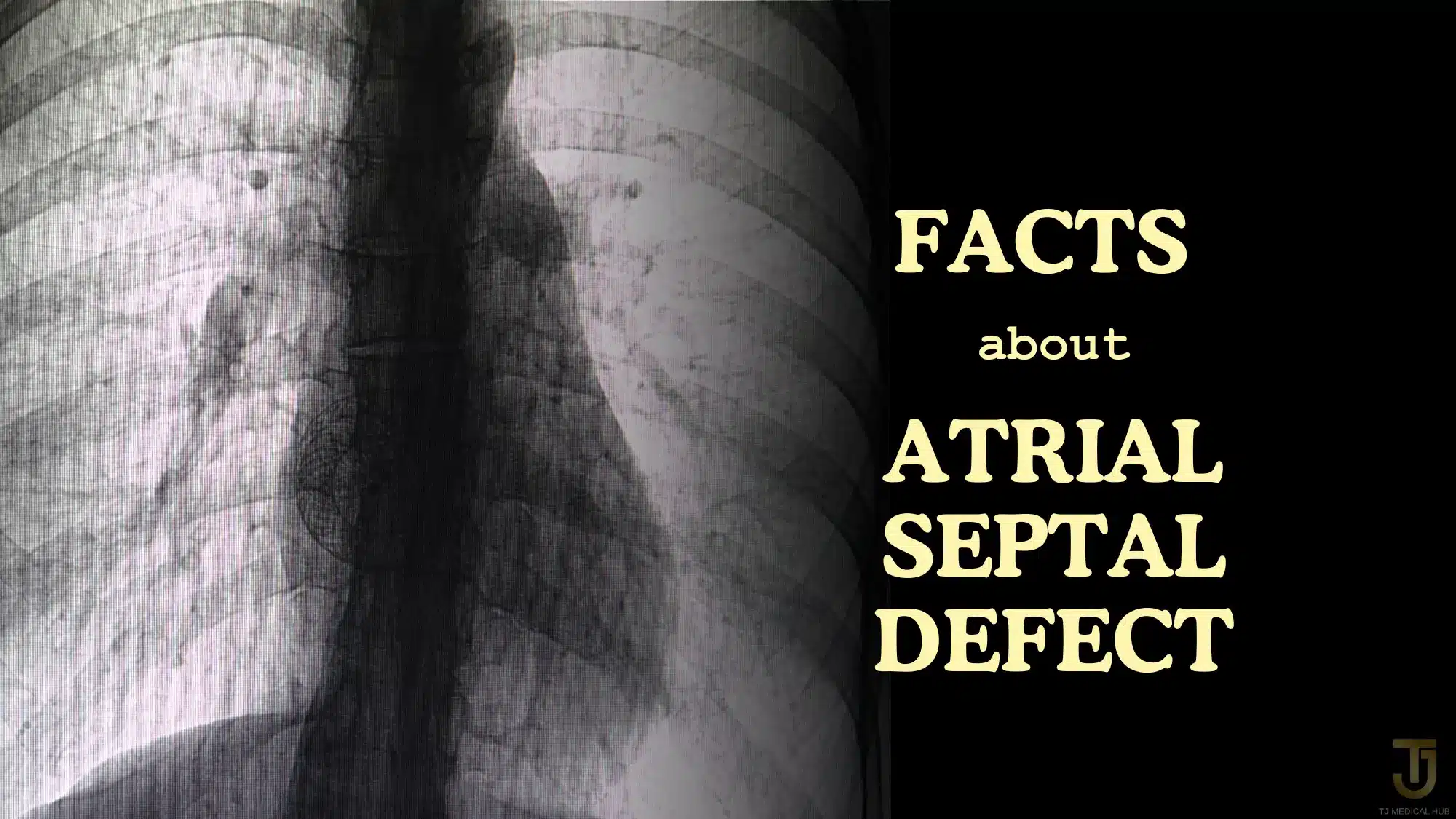
Transcatheter ASD Closure involves the insertion of metallic devices through a blood vessel in the leg, which are guided to the site of the hole in the heart wall. Once at the site, the device is released to cover the hole. Over the next 3 to 6 months, the body naturally develops tissue to cover the device. The choice of device depends on the characteristics and size of the ASD. This method offers the advantage of avoiding the risks and discomfort associated with open-heart surgery. The incisions are small, the recovery time is relatively short (approximately 48 hours), and patients tend to recuperate quickly. After the procedure, patients undergo follow-up examinations with echocardiograms at regular intervals, including monthly, every 3 months, 6 months, and annually, as recommended by the heart specialist. The success of Transcatheter ASD Closure depends on the expertise and experience of the cardiac team, as well as the availability of modern equipment and facilities for catheter-based heart examinations, resulting in favorable treatment outcomes.
The Transcatheter Closure of Secundum-Type Atrial Septal Defect (ASD) in Adults Using Special ASD Occluder Devices is Highly Successful with Minimal Complications.
After Birth
Atrial Septal Defects are typically present at birth, often showing no immediate signs. However, untreated or large defects may lead to symptoms such as:
- Frequent lung infections.
- Breathing difficulties.
- Fatigue during feeding (in infants).
- Shortness of breath during activity.
- Palpitations or awareness of heartbeat.
- Detectable heart murmur.
- Swelling in legs, feet, or abdomen.
- Stroke.
Diagnosis may not occur until adulthood, often through the identification of a murmur during a stethoscope examination. If signs or symptoms emerge, healthcare providers may order tests, with echocardiograms being the most common, to confirm the diagnosis. Echocardiograms use ultrasound to visualize the heart’s structure.
Treatment Options
The management of atrial septal defects hinges on factors like age at diagnosis, symptom severity, hole size, and associated conditions. Surgical intervention may be necessary for repair, while medications can alleviate symptoms, although no drug can close the hole itself.
In cases involving children, healthcare providers might initially monitor the defect to observe if spontaneous closure occurs. During this period, symptoms can be managed with medication. For large defects, closure may be recommended to prevent future complications, even if symptoms are mild. Closure can take place through cardiac catheterization or open-heart surgery. Post-procedure follow-up varies based on defect size, age, and any additional congenital conditions.
The transition from a hub to a local service is typically part of a broader effort to enhance the accessibility and quality of support and services. It aims to better align support with the specific needs and circumstances of the people it serves, promoting a more community-based and person-centered approach to care and wellbeing.
Nevertheless
Using a minimally invasive technique with special devices to close Secundum-type Atrial Septal Defect (ASD) in the heart isn’t suitable for every case. The feasibility depends on the size, anatomical characteristics, and the specific condition of the ASD in each patient. For instance, cases with ASDs larger than 36 millimeters, multiple ASDs, or patients with concomitant heart conditions, such as mitral valve regurgitation, should consider consulting a thoracic surgeon for further treatment via surgery. In Thailand, there are reputable hospitals and highly skilled doctors specializing in heart disease treatment. If you’re looking for expert guidance or considering treatment, please don’t hesitate to get in touch with TJ Medical Hub. We’re here to assist you.
